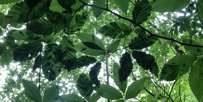
Recently I posted a blog on a paper by Paulo Vieira et al., reporting how the nematode Litylenchus crenatae subsp. mccannii (Lcm) [causal agent of beech leaf disease (BLD)] distorts the leaves of affected American beech trees (Fagus grandifolia). Now Leila Rose Fletcher and her colleagues have confirmed these structural changes and discussed how they might affect the anatomy and physiology of the leaves and harm the tree. For the full details, see this 2024 publication, cited at the end of this blog. Both articles contain stunning photographs of diseased leaf structures.
As Dr. Fletcher pointed out on a recent call involving most scientists and conservationists working on BLD, plant growth depends on the plant’s success in gaining more carbon (through photosynthesis) than it expends during growth, cell maintenance (respiration), and sugar storage. When plants open the stomata on their leaves to take in CO2 from the atmosphere, they lose water from the interior of their leaves.
Her team discovered that BLD-related leaf distortions reduced the tree’s carbon balance in two ways.
Two impacts of the nematode
First, alterations of the leaf structure reduce the tree’s photosynthetic rate (assimilation of carbon). The photosynthetic rate in affected leaves was 61% lower than in healthy leaves. The impact is heightened by the thinning of the beech tree’s canopy due to abortion of many leaf buds.
While veins in diseased leaves are also altered by BLD, the data in Fletcher et al. indicate that the main limitation on photosynthesis in symptomatic leaves is not from a decreased water supply, but from several limitations on stomatal exchange of CO2 with the atmosphere. First, stomata on symptomatic portions of diseased leaves are less dense, which means there are fewer openings through which CO2 can enter the leaf.
Second, the diseased leaves are thicker, meaning that once inside the leaf, CO2 molecules must travel farther from the stomatal pores to reach the photosynthetic cells. Fletcher et al. did not assess the possibility (raised in a separate study by Carta, et al.) that the stomata that are present are deformed, and that this might impact their function.
In addition, the deformed leaves demand more resources to grow and function. Production of the multiplicity of cells in affected portions of the leaf (these portions are 249% thicker than normal leaves) uses resources the tree would otherwise put into growth. In fact, the more severely symptomatic an individual leaf is, the more carbon the plant allocates to that leaf.Furthermore, the additional cell layers also appear to increase “operating costs” of these leaves, as seen in the higher respiration rate per unit leaf area. Finally, if the tree sheds deformed leaves and forms new ones, this further diverts resources.
Questions seeking answers
How is nematodes’ influence localized to domains bounded by second-order veins (large veins that branch off the central vein) – symptomatic and asymptomatic tissue in adjacent domains in the same leaf? Fletcher et al. propose that the presence of the nematode influences the physical or hormonal regulation of leaf development – but after the development of primary and secondary order veins (since they are not distorted). They place a high priority on investigating the tree’s hormonal signaling that might be disrupted by the nematode.
Given the carbon imbalance that the data in Fletcher et al. suggest might arise over time, will symptomatic trees face carbon shortages, and if so, will this eventually lead to mortality? Studies analyzing the non-structural carbohydrate (stored sugar) concentrations in symptomatic beech are urgently needed to explore this possibility.
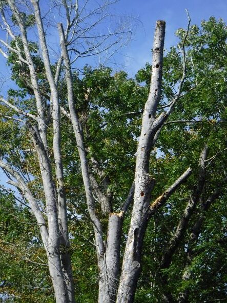
What is the impact of beech trees’ suboptimal vigor – short of mortality – on composition of plant communities and animals reliant on beech leaves and beechnuts? One possible causal factor raised by Fletcher et al. is reduced development of symbiotic relationships with ectomycorrhizal fungi, which can also reduce production of beech nuts. Dr. Fletcher concedes that there have been no studies yet of these possible effects. I add that many animals also depend on tree cavities – which are also common in beech trees.
SOURCES
Carta, L.K., S. Li, J. Mowery. 2023. Chapter 8 – Beech leaf disease (BLD), Litylenchus crenatae and its potential microbial virulence factors. In F.O. Asiegbu & A. Kovalchuck (Eds.), Forest microbiology Vol. 3 (PP. 183-192) Academic Press. https://doi.org/10.1016/B978-0-443-18694-3.00018-3
Fletcher, L.R. A.M. Borsuk, A.C. Fanton, K.M. Johnson, J. Richburg, J. Zailaa, C.R. Brodersen. 2024. Anatomical & physiological consequences of beech leaf disease in Fagus grandifolia L. Forest Pathoklogy. 2024;54:e12842 https://doi.org/10.1111.efp.12842
Vieira P., M.R. Kantor, A. Jansen, Z.A. Handoo, J.D. Eisenback. (2023) Cellular insights of beech leaf disease reveal abnormal ectopic cell division of symptomatic interveinal leaf areas. PLoS ONE October 5, 2023. 18(10) https://doi.org/10.1371/pone.0292588